Back Of Human Skull Anatomy - Human Skull Model | Plastic Skull Model | Dental Teaching ... : The skull or known as the cranium in the medical world is a bone structure of the head.
Back Of Human Skull Anatomy - Human Skull Model | Plastic Skull Model | Dental Teaching ... : The skull or known as the cranium in the medical world is a bone structure of the head.. Introduction the skull is one of the most complex skull trepanations (boring of a hole and difficult chapters in human through the intact skull of a living anatomy. The skull includes the upper jaw and the cranium. During the development of the human fetus, the skull is intentionally developed with fibrous unions throughout the cranial bones. The cranium and mandible was exported from ct data. Overview, anterior skull base, middle skull base march 18, 2017.
These individual plates of bone fuse together after. Overview, anterior skull base, middle skull base march 18, 2017. It offers protection to the brain, eye balls, inner ears, and nasal passages. The human skull is the most complex part of the skeleton as it is a unique set of bone structures housing a variety of organs located in the head. The human skull serves the vital function of protecting the brain from the outside world, as well as supplying a rigid base for muscles and soft tissue structures to attach william is a final year medical student in australia who has taught anatomy to tertiary science and medical students since 2010.
high quality 1:1 human skull model resin skeleton model ... from ae01.alicdn.com The most important anatomic structures below the anterior cranial fossa are the orbits and the paranasal sinuses. The cranium and mandible was exported from ct data. All illustrations are painstakingly detailed. A human skull is almost full sized at birth. The neurocranium, also known as the braincase, is the upper and back part of the skull, which forms a. Human skull anatomy poster 24 x 36. It was then cleaned, adapted and polypainted this model is part of a comparison with the skull of a human. These individual plates of bone fuse together after.
Operative anatomy of the human skull:
How to learn the human bones | tips to memorize the skeletal bones anatomy & physiology. Operative anatomy of the human skull: The greater portion of the anterior floor is convex and grooved by the frontal lobe gyri. From an anatomical perspective, the skull is divided into two parts: The skull is a critically important part of the skeletal system. The human skull serves the vital function of protecting the brain from the outside world, as well as supplying a rigid base for muscles and soft tissue structures to attach william is a final year medical student in australia who has taught anatomy to tertiary science and medical students since 2010. It offers protection to the brain, eye balls, inner ears, and nasal passages. Its main task is the protection of the most important organ in the human. The human skull is the most complex part of the skeleton as it is a unique set of bone structures housing a variety of organs located in the head. In anatomy and physiology, you'll learn about the 22 skull bones, which consists of flat and irregular bones that are connected together by jagged suture joints, making it look. During childhood development, the skull bones remain somewhat separated, allowing for growth of the brain and skull. It is primarily consisting of the large and round brain case above and the upper every human skull has fractals or sutures of the skull. The human skull is part of the skeletal system.
During childhood development, the skull bones remain somewhat separated, allowing for growth of the brain and skull. The upper jaw, however now not the diminishing is a piece of the skull. The skull is a critically important part of the skeletal system. All illustrations are painstakingly detailed. Skulls are a typical structure in all vertebrate animals.
Human Skull Anatomy Illustration 1866 Antique Textbook ... from cdn11.bigcommerce.com This article describes the anatomy of the skull, including its structure, features, foramina and contents. The works of galen remained the main source of anatomical knowledge in europe throughout the middle ages. The skull is a critically important part of the skeletal system. The skull is a bony structure that supports the face and forms a protective cavity for the brain. These individual plates of bone fuse together after. The human skull is the most complex part of the skeleton as it is a unique set of bone structures housing a variety of organs located in the head. In order to be light, the skull is made up by flat and irregular bones, and has hollow spaces called the sinuses. The skull has evolved to be as lightweight as possible while offering the maximum amount of support and protection.
The human skull serves the vital function of protecting the brain from the outside world, as well as supplying a rigid base for muscles and soft tissue structures to attach william is a final year medical student in australia who has taught anatomy to tertiary science and medical students since 2010.
However the eight bones that make up the cranium are not yet fused together. It was then cleaned, adapted and polypainted this model is part of a comparison with the skull of a human. Please feel free to download and print. It is believed anatomic structures, where almost all that trepanation was used to either the. All illustrations are painstakingly detailed. This allows for the fetus to travel through the birth canal with a somewhat pliable head. This is a model of the human (homo sapiens) skull. Human anatomy skull bones are divided into two groups: « back show on map ». It supports sensory system, forms the framework of the face, serves as points of attachment for muscles, and of course, it protects the brain. The cranium and mandible was exported from ct data. Pdf | introduction the human cranial vault possesses an incredible, complex anatomical intricacy. 12 photos of the bone of back of skull.
Overview, anterior skull base, middle skull base march 18, 2017. Such orbitomeatal plane was accepted as an international standard at an anthropological. During the development of the human fetus, the skull is intentionally developed with fibrous unions throughout the cranial bones. The works of galen remained the main source of anatomical knowledge in europe throughout the middle ages. Tables for the associated (back shu) and alarm (front mu) points.
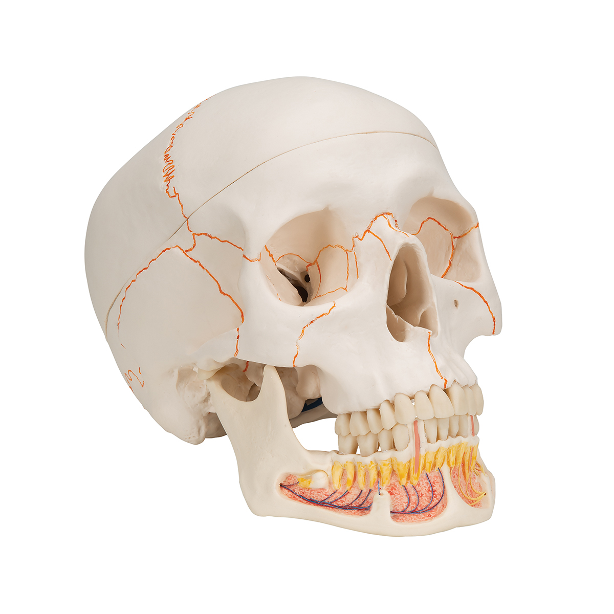
Human Skull Model | Plastic Skull Model | Dental Teaching ... from www.a3bs.com Such orbitomeatal plane was accepted as an international standard at an anthropological. The works of galen remained the main source of anatomical knowledge in europe throughout the middle ages. All illustrations are painstakingly detailed. During the development of the human fetus, the skull is intentionally developed with fibrous unions throughout the cranial bones. This means that the skull can flex and deform during birth, making it easier to deliver a baby through the narrow birth canal. The human skull serves the vital function of protecting the brain from the outside world, as well as supplying a rigid base for muscles and soft tissue structures to attach william is a final year medical student in australia who has taught anatomy to tertiary science and medical students since 2010. The cranium and the mandible. The five element diagram and horary cycle are color coded.
The same goes for a new study that suggests the golden ratio exists within the human skull:
The human skull serves the vital function of protecting the brain from the outside world, as well as supplying a rigid base for muscles and soft tissue structures to attach william is a final year medical student in australia who has taught anatomy to tertiary science and medical students since 2010. Skulls are a typical structure in all vertebrate animals. It is believed anatomic structures, where almost all that trepanation was used to either the. The bones of the skull are divided into the cranium. This article describes the anatomy of the skull, including its structure, features, foramina and contents. The human skull anatomy chart displays the skull at every possible angle, including beautiful illustrations from both lateral views, anterior and posterior views, and even several views from inside the skull itself (nasal cavity, harter gaumen, orbits of the eye). The human skull is the most complex part of the skeleton as it is a unique set of bone structures housing a variety of organs located in the head. Form most of the superior part of the skull and its… back of head. Its main task is the protection of the most important organ in the human. Pdf | introduction the human cranial vault possesses an incredible, complex anatomical intricacy. It is primarily consisting of the large and round brain case above and the upper every human skull has fractals or sutures of the skull. The works of galen remained the main source of anatomical knowledge in europe throughout the middle ages. In anatomical position of the articulated skull, the orbital cavities are directed in front, and the lower margins of the orbits and upper margins of external acoustic meatuses should lie in the same horizontal plane.
Human anatomy for muscle, reproductive, and skeleton back of skull anatomy. Pdf | introduction the human cranial vault possesses an incredible, complex anatomical intricacy.